

VLA Update July 2010
Johne's disease is becoming one of the most frequently reported enteric diseases in adult cattle, says the Veterinary Laboratory Agency's UK animal health survey.Reproductive Diseases
AbortionThirsk isolated Listeria monocytogenes from the foetal stomach content of a bovine foetus which had been aborted at 200 days gestation. It was the only abortion recorded from a group of 110 dairy cows. There was a history of the cows being fed silage and that was seen as the most likely origin of the infection.
Penrith diagnosed Chlamydophila abortus infection as the cause of bovine abortion in a herd where there had been 3 abortions in a group of 60 milking cows. At necropsy there was evidence of a severe, diffuse placentitis. The diagnosis was confirmed by PCR. Coxiella burnetti (Q fever) organisms were identified by Winchester in the placental material that accompanied a seven-month gestation aborted calf. The farm was currently experiencing an upsurge of abortions, but the organism had only been confirmed in a single submission. Appropriate public health advice was given to the farm staff.
Alimentary tract diseases
Idiopathic EnteritisPenrith diagnosed Idiopathic necrotising enteritis of suckler calves in a 2- month-old Limousin suckler calf; one of two similarly affected animals, both of which had developed ascites. Bury also diagnosed a case affecting a suckler calf two weeks after turnout. Necropsy revealed peritoneal fibrin tags, a raised ulcerated area in the rumen, distended distal jejunum and diphtheritic enteritis in the ileum. There were extensive well-defined areas of lung consolidation and a fibrinous pleurisy.
SalmonellosisLeahurst isolated Salmonella Dublin from two 3-month-old Aberdeen Angus x calves from a group of 29 introduced from a neighbouring farm a month earlier. One had died on the farm of origin and one other died after they were moved.
Langford diagnosed Salmonellosis involving a number of different serotypes. Salmonella Dublin was identified as the cause of enteritis and pneumonia affecting three out of 120, eight to ten week old bull calves one of which died. Salmonella Typhimurium phage type 193 was the cause of malaise and diarrhoea affecting yearling animals whilst Salmonella Mbandaka was the cause of neonatal diarrhoea in Holstein Friesian heifer calves on a central heifer rearing unit serving four or five dairy herds. In another case Salmonella Anatum was associated with sudden milk drop four months post calving, anorexia and scouring in a single Holstein Friesian dairy cow.
Starcross diagnosed Fasciolosis (liver fluke) on seven farms and reported cases of rumen fluke (Paramphistomum) on three of them. Other co infections included Salmonella Dublin (2 farms) and Johne’s disease (1 farm).
Langford also diagnosed fasciolosis in two adult cows with a history of diarrhoea and weight loss.
Both Preston and Langford reported Johne’s disease to be the most frequent diagnosis of enteric disease in adult cattle. The majority of diagnoses were based on serology and cases affected both dairy and beef cattle. Diarrhoea, weight loss and milk drop were the most commonly described clinical signs.
Fatty liver SyndromeA section of liver was submitted to Langford from a heifer with a history of acute onset jaundice and death. It was one of a group of cattle which had recently been imported from Europe. A field post mortem examination by the attending veterinarian revealed a swollen and discoloured liver. Histopathological examination at VLA revealed marked fatty change and hepatocellular degeneration with irregular foci of cell necrosis and collapse with mild inflammation, indicative of acute degenerative hepatopathy. This suggested an underlying metabolic or toxic injury to the liver. Copper levels were measured and were found to be within the reference range. No specific cause of the acute deterioration and necrosis of the liver was identified at the time but subsequently further animals also presented similarly within the same group and other groups of imported cattle. These animals may be subjected to metabolic stress during the process of importation and acclimatisation to new management systems within the UK and this could have resulted in the changes observed.
Respiratory Diseases
Histophilus SomniPenrith diagnosed Histophilus somni pneumonia in a 7-week-old Belgian Blue calf, the only one affected in a group of 10. At post-mortem the animal showed dramatic uniform consolidation of all lung lobes with a typical cranio-ventral distribution and mucopurulent material in the airways, findings that were considered typical of Histophilus pneumonia.
IBRFour yearling cattle were turned out to grass following administration of worming boluses. Within 48 hours all four animals were found dead. Post mortem examination of an affected animal at Thirsk revealed severe diphtheresis along the entire length of the trachea and consolidation of approximately 50 per cent of lung parenchyma. Fluorescent antibody testing confirmed the presence of IBR virus within tracheal cells and Mannheimia species were cultured from the lung tissue. Although the postmortem showed no evidence that the animal had been roughly handled, with no bruising to the carcase or oesophagus, it was considered likely that the bolusing of the four animals in addition to turnout was a stressful event which may have triggered latent IBR infection. Langford also diagnosed fasciolosis in two adult cows with a history of diarrhoea and weight loss.
Lungworm (Dictyocaulosis)Thirsk diagnosed an outbreak of lungworm in the first week of July, with 70 from a group of 90 beef finisher calves affected with coughing and weight loss. At necropsy severe lungworm infestation, with secondary Pasteurella multocida infection, was demonstrated in one of the submitted animals. The calves were not vaccinated against husk. Leahurst also diagnosed a case affecting a 23-month-old Hereford bull which died after being ill with pyrexia, severe dyspnoea and coughing for 2 days. There was no response to treatment with antibiotics. An incidental finding in this bull was the presence in the rumen and reticulum of large numbers of very small shot, which had been flattened. It seemed likely that this was ‘rat shot’ or ‘dust-shot’ which might have been used to control vermin around a feed mill on the bull’s farm of origin and might have accidentally entered the mill. The lead in such material is usually relatively unavailable biologically but metallic objects can remain in the forestomachs for a period of years and can become more bioavailable in the presence of acidosis. The kidney lead level of this bull was very slightly elevated above expected background level.
Other Diseases
BabesiosisPreston diagnosed Babesiosis affecting 8 out of group of 100 dairy cows. Presenting signs were haematuria, pyrexia, pale mucous membranes, tachycardia, weakness and anorexia. All the affected cows were found to be anaemic and Babesia species were identified on examination of blood smears.
Penrith also diagnosed the disease following examination of a blood sample from an adult cow. They remarked that the condition occurs sporadically in Cumbria but is restricted to several relatively well known areas.
Thirsk diagnosed Clostridial myositis, caused by Clostridium chauvoei, (blackleg) as a cause of death in three animals from a group of 16, one-year-old British Blue cross beef finisher calves. Post-mortem of one affected animal revealed typical lesions in the deep muscles of the neck and the muscles covering the rib cage as well as the intercostal muscles and parts of the lungs and the heart. Preston also diagnosed blackleg affecting a single 3 ½ year old Swedish red and white dairy cow from a herd of 240 which had been presented for post mortem examination having calved one week earlier.
Staphylococcal DermatitisSkin scrapes and hair samples were submitted to Sutton Bonnington from a group of two-year-old dairy heifers to investigate a severe outbreak of pruritus and exudative dermatitis. Approximately 50 per cent of the group were affected with the primary site of localisation being over the withers. Routine examination for parasites and dermatophytes was unremarkable with the only consistent finding being the recovery of an α-β-haemolytic Staphylococcus aureus. Response to antibiotics was achieved in conjunction with changing the current feeding regime as the animals were being fed through a barrier and it was felt that, even though the barrier had no abrasive edges, it acted as a focal point for the collection of scab debris, so allowing transfer to unaffected animals. The source of the infection was not clear as the group was a stable one with no introduction of new animals and no changes in their management for some time. An external source of infection was proposed but not identified.
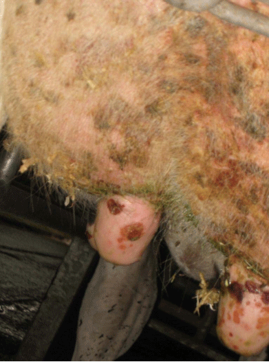
Bovine herpes mammilitisA high convalescent SNT titre to bovine herpes mammillitis was confirmed in a 6th lactation Holstein/Friesian cow showing widespread vesicular coalescing and granulating lesions over the udder (Fig 1), it was the only animal affected. No viral particles were demonstrated by electron microscopy.
Figure 1
Bovine Herpes Mammilitis


