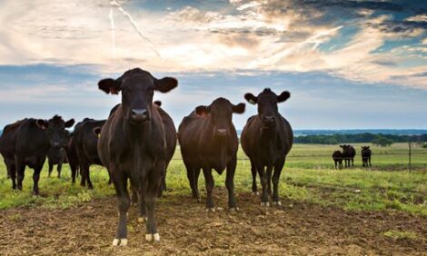
Lameness in Newly Arrived Feedlot Calves
Lameness affects performance, animal welfare (in the feedlot and in transport) and economics. The incidence of lameness varies, and different types of lameness have different causes (e.g. genetic predisposition, frostbitten feet, footrot, nutrition, mycoplasma, injury, etc.). Research by the Canadian beef industry is investigating P3 necrosis in feedlot cattle. 8 April 2012
 2 minute read
What is P3 necrosis
P3 necrosis is a specific type of lameness that affects the outside claw of the hindfoot, and typically occurs within one to seven days after arriving at the feedlot.
The coronary band at the top of the hoof splits, and the hoof wall often sloughs off after two to four weeks. Very excitable cattle may be more prone to this condition when they damage the soles of their feet while struggling in the chute, but cause and effect may be difficult to separate.
It is unclear whether struggling causes the initial injury, or if animals struggle because the foot is already in pain. The disease is sporadic, but tends to cluster by truckload and feedlot pen.
Extreme lameness results in a rapid loss in body condition and poor performance. Approximately half of the animals will recover; the remainder are euthanized because of poor performance and concerns about animal welfare.
What the research will look at?
1. Work with feedlot veterinary practices to better understand which cattle are affected (gender and age), number of days on feed when P3 necrosis appears, seasonality, clustering by farm of origin, trailer load, etc.
2. The same feedlot veterinarians will submit feet from cattle euthanized due to lameness, as well as other causes to see if the location and severity of the lesion will provide insights into possible causes. The veterinarians will record the exact location of hoof lesions in lame cattle, and conduct biomechanical testing (hoof hardness, hoof moisture content, etc.) of affected and unaffected claws.
3. The hooves will also be used for micronutrient analysis to determine if inadequate nutrition contributes to a softer claw, making it more susceptible to injury. Potassium, calcium, phosphorus, sodium, magnesium, manganese, sulfur, copper, zinc and biotin levels will be measured in normal and affected hooves to see if particular nutritional deficiencies predispose the hoof to developing the P3 lesion.
4. Normal and affected hooves from dead cattle will be examined using ultrasound, computed tomography, X-rays, nuclear scintigraphy and the biomedical beamline of the Canadian Light Source to examine structural differences. Live cattle will also be imaged to compare circulation in the hooves of normal cattle and those with P3 necrosis.
5. Microbiological testing will look for infectious pathogens like Treponema bacteria (which is involved in digital dermatitis in dairy cattle) and bovine viral diarrhea virus.
Implications
A better understanding of P3 necrosis is important to identify strategies that may effectively prevent or control it.
April 2012